

Muscles That Elevate The Mandible
The muscles of mastication are a group of muscles responsible for chewing (i.e. motion of the mandible at the temporomandibular articulation). These muscles originate from the surface of the skull and insert onto the mandible.¹
There are four muscles that contain the muscles of mastication, including masseter, temporalis, lateral pterygoid and medial pterygoid.¹
This article will explore the origin, insertion, action, innervation and blood supply of the masticatory muscles. Sure pathologies associated with detail muscles of mastication will likewise be discussed.¹
You might also be interested in our Anatomy Flashcard Collection which contains over 2000 beefcake flashcards in addition to avant-garde features such as spaced repetition. 🫁
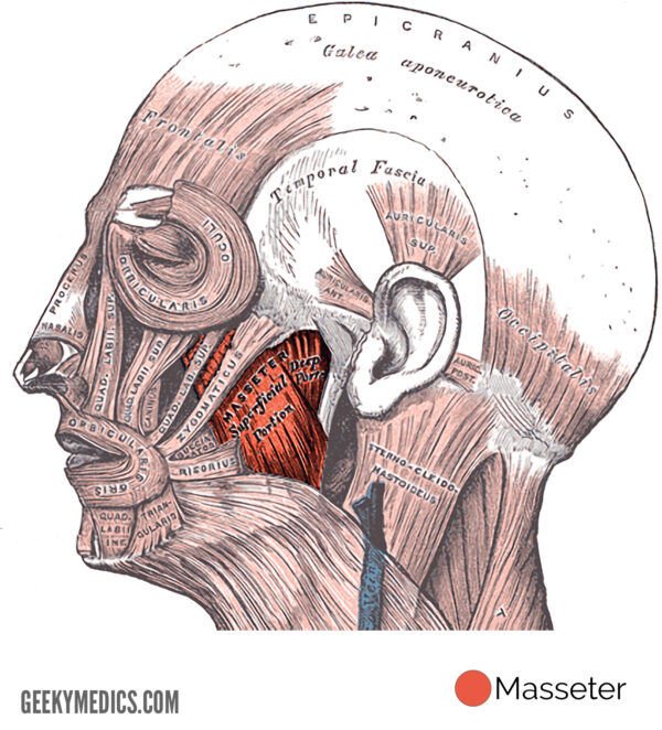
Masseter
- This musculus composes of two main parts: the deep and superficial heads.¹
- This musculus is only found in mammals.
Origin
- The masseter originates at the zygomatic curvation and maxillary procedure of the zygomatic bone.
Insertion
- The insertion site is located at the lateral surface of the ramus and angle of the mandible.¹
Action
- Bilateral wrinkle volition beetle and elevate the mandible.¹
- Unilateralwrinkle will cause a pocket-size amount of side-to-side movement (lateral deviation).
Innervation
- The mandibular division of the trigeminal nerve (V3).¹
Blood supply
- The masseteric artery (a branch of the 2nd part of the maxillary artery),¹ facial artery and the transverse facial co-operative of the superficial temporal artery.
Clinical relevance
Bilateral enlargement of this muscle can occur in patients who frequently grind their teeth (also known every bit bruxism).
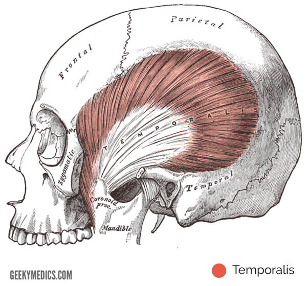
Temporalis
- This large, fan-shaped muscle is located on the lateral aspect of the skull.
- It occupies the temporal fossa.
Origin
- The temporalis originates at the temporal fossa upwardly to the inferior temporal line and the zygomatic bone.
Insertion
- The coronoid process and ramus of the mandible.¹
Activity
- Bilateral wrinkle of the temporalis muscle contributes to retraction and elevation of the mandible.
- Unilateralcontraction will crusade a small amount of side-to-side motion (lateral divergence).¹
Innervation
- The mandibular sectionalization of the trigeminal nerve (V3).¹
Claret supply
- The deep temporal arteries, which are branches of the 2nd part of the maxillary artery.¹
Clinical relevance
Hurting in the temporalis muscle tin be a symptom of bruxism. Additionally, rupture of the myotendinous insertion of the temporalis tin occur due to farthermost teeth clenching (e.g. during seizures).
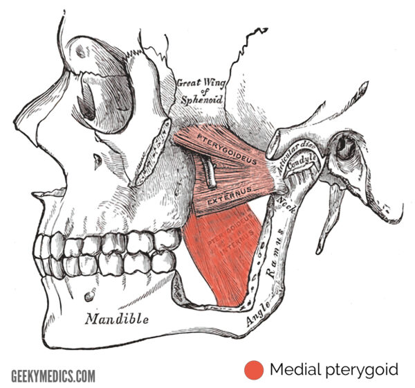
Medial pterygoid
- This musculus is separated into deep and superficial sections.¹
Origin
-
-
- The deep head originates from the medial attribute of the lateral pterygoid plate of the sphenoid.
- The superficial caput originates from the pyramidal process of the palatine bone and maxillary tuberosity.¹
Insertion
-
-
- Both the deep and superficial heads insert onto the medial surface of the ramus and angle of the mandible.
- The insertion of the medial pterygoid forms a tendinous band with the insertion of the masseter called the pterygo-masseteric sling.¹
-
Activity
-
-
- Bilateral contraction of this muscle elevates and protrudes the jaw.
- Unilateral contraction volition deviate the mandible to the contralateral side.
-
Innervation
-
-
- The mandibular partition of the trigeminal nerve (V3).¹
-
Claret supply
-
-
- The pterygoid branches of the maxillary avenue
-
Clinical relevance
- Limitation of jaw opening (also known as trismus) sometimes occurs as a issue of radiotherapy in the treatment of head and neck cancers.
- This occurs due to fibrosis of the pterygo-masseteric sling.
Medial pterygoid
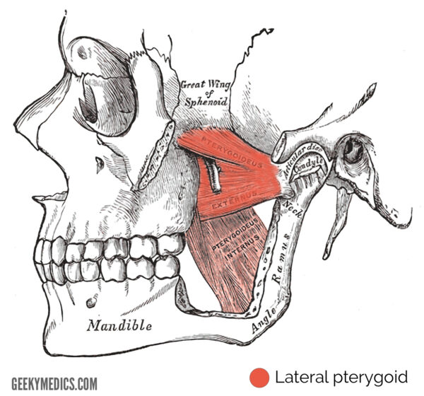
Lateral pterygoid
-
-
- This muscle lies mostly superficial to the medial pterygoid and comprises of superior and inferior sections.¹
-
Origin
-
-
- The superior head originates from the infratemporal surface of the greater fly of the sphenoid, whilst the inferior head originates from the lateral surface of the lateral pterygoid plate of the sphenoid.
-
Insertion
-
-
- The fibres from both heads converge to adhere to the pterygoid fovea on the cervix of the mandible.
- A portion of the superior head attaches to the capsule and articular disc of the temporomandibular joint.¹
-
Action
-
-
- Bilateral contraction pulls the mandibular condyle and articular disc anteriorly (i.e. protrusion). This movement is thought to be integral to the opening of the jaw.
- Unilateral contraction volition deviate the mandible to the contralateral side.
-
Innervation
-
-
- The mandibular division of trigeminal nervus (V3).¹
-
Claret supply
-
-
- The pterygoid branches of the maxillary artery.¹
-
Lateral pterygoid
Reviewer
Dr James Nott
Anatomy Lecturer & Anatomy Pb at Geeky Medics
References
-
-
- Drake, R. L., Wayne Vogl, A., Mitchell, A. (2009). Greyness'south anatomy for students second edition. Elsevier health sciences.
-
-


Muscles That Elevate The Mandible,
Source: https://geekymedics.com/muscles-of-mastication/
Posted by: cannonbenty1991.blogspot.com
0 Response to "Muscles That Elevate The Mandible"
Post a Comment